Suspected malignant skin excisions: Difference between revisions
Jump to navigation
Jump to search
+Conditions |
|||
| Line 28: | Line 28: | ||
{|class="wikitable" | {|class="wikitable" | ||
|- | |- | ||
! Condition<ref name="PaolinoDonati2017">Initially copied from: {{cite journal|last1=Paolino|first1=Giovanni|last2=Donati|first2=Michele|last3=Didona|first3=Dario|last4=Mercuri|first4=Santo|last5=Cantisani|first5=Carmen|title=Histology of Non-Melanoma Skin Cancers: An Update|journal=Biomedicines|volume=5|issue=4|year=2017|pages=71|issn=2227-9059|doi=10.3390/biomedicines5040071}}</ref> - "This article is an open access article distributed under the terms and conditions of the Creative Commons Attribution (CC BY) license (http://creativecommons.org/licenses/by/4.0/)." !! Characteristics<ref name="PaolinoDonati2017"/> !! | ! Condition<ref name="PaolinoDonati2017">Initially copied from: {{cite journal|last1=Paolino|first1=Giovanni|last2=Donati|first2=Michele|last3=Didona|first3=Dario|last4=Mercuri|first4=Santo|last5=Cantisani|first5=Carmen|title=Histology of Non-Melanoma Skin Cancers: An Update|journal=Biomedicines|volume=5|issue=4|year=2017|pages=71|issn=2227-9059|doi=10.3390/biomedicines5040071}}</ref> - "This article is an open access article distributed under the terms and conditions of the Creative Commons Attribution (CC BY) license (http://creativecommons.org/licenses/by/4.0/)."</ref> !! Characteristics<ref name="PaolinoDonati2017"/> !! | ||
|- | |- | ||
| [[Actinic keratosis]] || Atypical keratinocytes confined on basal layer. | | [[Actinic keratosis]] || Atypical keratinocytes confined on basal layer. | ||
| Line 36: | Line 36: | ||
| Keratoacanthoma || Symmetrical and circumscribed proliferation of keratinocytes, with central horn plug, with epidermis that extends over the tumor. Highly differentiated SCC. | | Keratoacanthoma || Symmetrical and circumscribed proliferation of keratinocytes, with central horn plug, with epidermis that extends over the tumor. Highly differentiated SCC. | ||
|- | |- | ||
| Invasive squamous cell carcinoma || Atypical and pleomorphic keratinocytes, involving the dermis and the sub-cutis with a potential metastatic spread. | | Invasive [[squamous cell carcinoma of the skin]] || Atypical and pleomorphic keratinocytes, involving the dermis and the sub-cutis with a potential metastatic spread. | ||
|- | |- | ||
| Queyrat’s erytroplasia || As Bowen, but in the mucosa. | | Queyrat’s erytroplasia || As Bowen, but in the mucosa. | ||
Revision as of 10:40, 23 October 2019
Author:
Mikael Häggström [note 1]
Fixation
- Generally: Buffered 4% formaldehyde.[1]
Common targets
If directly suspected from the referral, see:
Gross evaluation
Note:
- Color
-
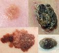 See separate article in case of dark skin focalities
- Well-defined or diffuse border
- Size
- Any elevation
Tissue selection


In table above, each top image shows recommended lines for cutting out slices to be submitted for further processing. Bottom image shows which side of the slice that should be put to microtomy. Dashed lines here mean that either side could be used. Further information: Gross processing of skin excisions
Microscopic evaluation
Look for atypical cells. If found, look for:
- Any pigmentation: Evaluate as dark skin focalities.
- Similarity to squamous cells: See below:
Squamous cell-like proliferations
| Condition[3] - "This article is an open access article distributed under the terms and conditions of the Creative Commons Attribution (CC BY) license (http://creativecommons.org/licenses/by/4.0/)."</ref> | Characteristics[3] | |
|---|---|---|
| Actinic keratosis | Atypical keratinocytes confined on basal layer. | |
| Bowen | Atypical keratinocytes at every layer of epidermis. | |
| Keratoacanthoma | Symmetrical and circumscribed proliferation of keratinocytes, with central horn plug, with epidermis that extends over the tumor. Highly differentiated SCC. | |
| Invasive squamous cell carcinoma of the skin | Atypical and pleomorphic keratinocytes, involving the dermis and the sub-cutis with a potential metastatic spread. | |
| Queyrat’s erytroplasia | As Bowen, but in the mucosa. | |
| Adnexal carcinomas | Squamous differentiation, but does not show connection with the epidermis and highlights adnexal features. | |
| Adenosquamous carcinoma | Mixed glandular and squamous differentiation. | |
| Verrucous squamous cell carcinoma[notes 1] | Exophytic squamous proliferation with marked papillomatosis and low atypia and the presence of koilocyte-like changes | |
| Epithelioma cuniculatum | Squamous cell carcinomaof the foot. Histologically is characterized hyperkeratosis, acanthosis with an undulating, densely keratinized, well differentiated squamous epithelium, deeply penetrating the soft tissues. | |
| Inverted follicular keratosis | Sharply circumscribed endophytic verrucous proliferation with prominent squamous features. | |
| Seborrheic keratosis | Acanthosis, absence of atypia, pseudo-horn cysts, in inflamed lesions, mitoses may be present. | |
| Bowenoid papulosis | Atypical keratinocytes and mitoses. Histology similar to Bowen’s disease. | |
| Metastasis | Personal medical history of the patient, nodular proliferation without connection to epidermis, immunohistochemical evaluation. |
Notes
- ↑ When present in ano-genital region, verrucous squamous cell carcinoma is also known with the term of Buschke–Löwenstein tumor
- ↑ For a full list of contributors, see article history. Creators of images are attributed at the image description pages, seen by clicking on the images. See Patholines:Authorship for details.
- ↑ The excision examples show a normal mole (upper row, benign appearance) and a superficial basal cell carcinoma (lower row, suspected malignancy).
Main page
References
- ↑ Katarzyna Lundmark, Krynitz, Ismini Vassilaki, Lena Mölne, Annika Ternesten Bratel. Handläggning av hudprover – provtagningsanvisningar, utskärningsprinciper och snittning (Handling of skin samples - Instructions for sampling, cutting and incision. KVAST (Swedish Society of Pathology). Retrieved on 2019-09-09.
- ↑ There are many variants for the processing of skin excisions. These examples use aspects from the following sources:
- . Handläggning av hudprover – provtagningsanvisningar, utskärningsprinciper och snittning (Handling of skin samples - sampling instructions, cutting principles and incision. Swedish Society of Pathology.
- For number of slices and coverage of lesions, depending on size. - Monica Dahlgren, Janne Malina, Anna Måsbäck, Otto Ljungberg. Stora utskärningen. KVAST (Swedish Society of Pathology). Retrieved on 2019-09-26.
- For slices towards the tips to determine radicality, which can be parallel to the slices through the lesions (shown), or as longitudinal slices that go through each tip. - . Dermatopathology Grossing Guidelines. University of California, Los Angeles. Retrieved on 2019-10-23.
- For microtomy of the most central side at the lesion - "The principles of mohs micrographic surgery for cutaneous neoplasia
- With a "standard histologic examination" that, in addition to the lesion, only includes one section from each side along the longest diameter of the specimen.
- It also shows an example of circular coverage, with equal coverage distance in all four directions.
- The entire specimen may be submitted if the risk of malignancy is high. - . Handläggning av hudprover – provtagningsanvisningar, utskärningsprinciper och snittning (Handling of skin samples - sampling instructions, cutting principles and incision. Swedish Society of Pathology.
- ↑ 3.0 3.1 Initially copied from: Paolino, Giovanni; Donati, Michele; Didona, Dario; Mercuri, Santo; Cantisani, Carmen (2017). "Histology of Non-Melanoma Skin Cancers: An Update ". Biomedicines 5 (4): 71. doi:. ISSN 2227-9059.
Image sources