Cervical cytology: Difference between revisions
Jump to navigation
Jump to search
→Dysplasia: SILs Tags: Mobile edit Mobile web edit |
→Adequacy: Calculation |
||
| Line 4: | Line 4: | ||
}} | }} | ||
==Adequacy== | ==Adequacy== | ||
Adequacy should always be stated, either as "Satisfactory" or "Unsatisfactory". For estimating the number of cells, determine the following: | |||
*The area of your field of view at high power (see the [[Evaluation]] chapter) | |||
*The total size of the relevant area on the microscope slide. | |||
*Look at 10 representative high power fields (HPFs) within that area, and calculate the average number of cells per high power field. | |||
{| | |||
|valign=center| Total number of cells = Average number of cells per HPF * || <u>Total size of area</u><br>HPF area | |||
|} | |||
Also state whether the endocervical/transformation zone is present or absent. Count an endocervical component as present if there are 10 or more endocervical or squamous metaplastic cells.<ref name="CibasDucatman">{{cite book | last=Cibas | first=Edmund S. | last2=Ducatman | first2=Barbara S. | title=Cytology : diagnostic principles and clinical correlates | publication-place=Philadelphia, PA | date=2021 | isbn=978-0-323-63637-7 | oclc=1138033641 | page=9}}</ref> | Also state whether the endocervical/transformation zone is present or absent. Count an endocervical component as present if there are 10 or more endocervical or squamous metaplastic cells.<ref name="CibasDucatman">{{cite book | last=Cibas | first=Edmund S. | last2=Ducatman | first2=Barbara S. | title=Cytology : diagnostic principles and clinical correlates | publication-place=Philadelphia, PA | date=2021 | isbn=978-0-323-63637-7 | oclc=1138033641 | page=9}}</ref> | ||
Revision as of 19:32, 15 August 2022
Author:
Mikael Häggström [note 1]
Adequacy
Adequacy should always be stated, either as "Satisfactory" or "Unsatisfactory". For estimating the number of cells, determine the following:
- The area of your field of view at high power (see the Evaluation chapter)
- The total size of the relevant area on the microscope slide.
- Look at 10 representative high power fields (HPFs) within that area, and calculate the average number of cells per high power field.
| Total number of cells = Average number of cells per HPF * | Total size of area HPF area |
Also state whether the endocervical/transformation zone is present or absent. Count an endocervical component as present if there are 10 or more endocervical or squamous metaplastic cells.[1]
Very common findings
-
Bacterial flora (usually rod-shaped Lactobacilli), does not need reporting
Main conditions to exclude or confirm
Squamous atypia, seen mainly as cells with increased nucleus/cytoplasm ratio, nuclear hyperchromasia and irregular nuclear outline.
-
Atypical squamous cells of undetermined significance (ASCUS), with only few slightly atypical cells
-
Low-grade squamous intraepithelial lesion (LSIL)
-
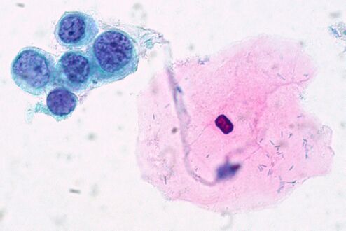 High-grade squamous intraepithelial lesion (HSIL)


_(9392112063).jpg)
.jpg)
Notes
- ↑ For a full list of contributors, see article history. Creators of images are attributed at the image description pages, seen by clicking on the images. See Patholines:Authorship for details.
Main page
References
- ↑ Cibas, Edmund S.; Ducatman, Barbara S. (2021). Cytology : diagnostic principles and clinical correlates . Philadelphia, PA. p. 9. ISBN 978-0-323-63637-7. OCLC 1138033641.
Image sources