
Invasive squamous cell carcinoma of the cervix
Author:
Mikael Häggström [note 1]
Microscopic evaluation
Infiltration as irregular anastomosing nests or single cells.[1] In contrast to tangentially cut projections of glands involved by HSIL, the invasive nests of tumor cells are irregular and accompanied by a distinct stromal reaction of edema and chronic inflammatory cells.
-
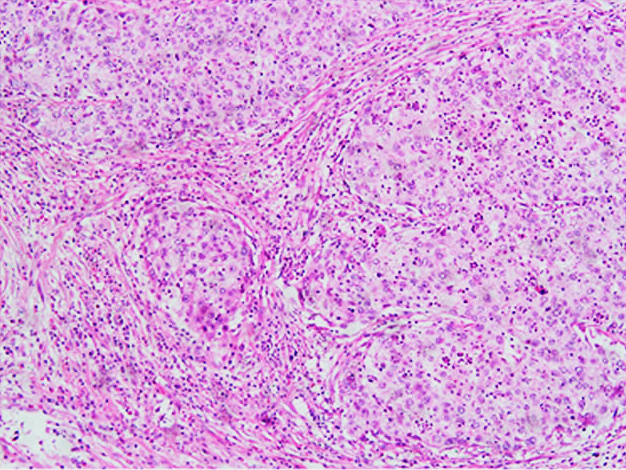 Invasive SCC of the cervix
 -
![Invasive SCC of the cervix[image 1]](https://upload.wikimedia.org/wikipedia/commons/a/a5/Invasive_Squamous_Carcinoma_of_the_Cervix._Cold_Knife_Cone_(6076190079).jpg) Invasive SCC of the cervix[image 1]
Invasive SCC of the cervix[image 1]
Staging
As per AJCC 9:[2]
| T CATEGORY | FIGO STAGE | T CRITERIA |
|---|---|---|
| TX | Primary tumor cannot be assessed | |
| T0 | No evidence of primary tumor | |
| T1 | I | Carcinoma is strictly confined to the cervix (extension to the corpus should be disregarded) |
| T1a | IA | Invasive carcinoma that can be diagnosed only by microscopy with maximum depth of invasion ≤5 mm |
| T1a1 | IA1 | Measured stromal invasion ≤3 mm in depth |
| T1a2 | IA2 | Measured stromal invasion >3 mm and ≤5 mm in depth |
| T1b | IB | Invasive carcinoma with measured deepest invasion >5 mm (greater than stage IA); lesion limited to the cervix uteri with size measured by maximum tumor diameter; note: the involvement of vascular/lymphatic spaces should not change the staging, and the lateral extent of the lesion is no longer considered |
| T1b1 | IB1 | Invasive carcinoma >5 mm depth of stromal invasion and ≤2 cm in greatest dimension |
| T1b2 | IB2 | Invasive carcinoma >2 cm and ≤4 cm in greatest dimension |
| T1b3 | IB3 | Invasive carcinoma >4 cm in greatest dimension |
| T2 | II | Carcinoma invades beyond the uterus but has not extended onto the lower one-third of the vagina or to the pelvic wall |
| T2a | IIA | Involvement limited to the upper two-thirds of the vagina without parametrial invasion |
| T2a1 | IIA1 | Invasive carcinoma ≤4 cm in greatest dimension |
| T2a2 | IIA2 | Invasive carcinoma >4 cm in greatest dimension |
| T2b | IIB | With parametrial invasion but not up to the pelvic wall |
| T3 | III | Carcinoma involves the lower one-third of the vagina and/or extends to the pelvic wall and/or causes hydronephrosis or nonfunctioning kidney; note: the pelvic wall is defined as the muscle, fascia, neurovascular structures, and skeletal portions of the bony pelvis; cases with no cancer-free space between the tumor and pelvic wall by rectal examination are FIGO stage III |
| T3a | IIIA | Carcinoma involves the lower one-third of the vagina, with no extension to the pelvic wall |
| T3b | IIIB | Extension to the pelvic wall and/or hydronephrosis or nonfunctioning kidney (unless known to be due to another cause) |
| T4 | IVA | Carcinoma has involved (biopsy-proven) the mucosa of the bladder or rectum or has spread to adjacent organs (bullous edema, as such, does not permit a case to be assigned to stage IVA) |
Microscopy report
Example:
| Cervix, uterus, bilateral tubes, ovaries and parametria, hysterectomy and bilateral salpingo-oophorectomy:
SYNOPTIC REPORT:
|
Notes
- ↑ For a full list of contributors, see article history. Creators of images are attributed at the image description pages, seen by clicking on the images. See Patholines:Authorship for details.
Main page
References
- ↑ Author: Gulisa Turashvili, M.D., Ph.D.. Cervix - Squamous cell carcinoma and variants. Pathology Outlines. Last author update: 24 September 2020. Last staff update: 13 December 2022
- ↑ Olawaiye AB, Baker TP, Washington MK, Mutch DG (2021). "The new (Version 9) American Joint Committee on Cancer tumor, node, metastasis staging for cervical cancer. ". CA Cancer J Clin 71 (4): 287-298. doi:. PMID 33784415. Archived from the original. .
.jpg)